 |
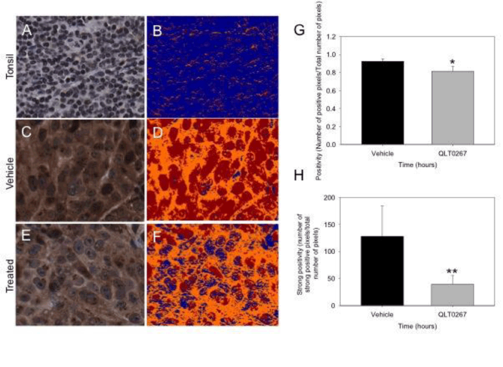
| Figure 8: GSK-3 is activated by QLT0267 in vivo. Vehicle and QLT0267 treated tumor cores were subject to pGSK-3 staining and quantification using Aperio Image Scope software. Tonsil tissue exhibits very little moderate intensity staining (A and B). The digital images of Vehicle and QLT0267 cores indicate reduced pGSK-3 in QLT0267 treated tumor cores (E and F) as compared to vehicle treated samples (C and D). The colorized mark–up image shows different levels of pixel intensity where blue represents negative pixels, orange moderately intense positive pixels and red strongly intense positive staining. Strong staining (red) is decreased in the tumor cores treated with QLT0267 (E and F). Quantification of overall positivity (G), and strongly positive pixels based on higher threshold (H) was performed using the AperioImageScope software analysis. QLT0267 treated tumors show a significant decrease in positivity (P=0.0089) and strongly positive (P=0.0113) pixels compared to vehicle treated tumors. |