 |
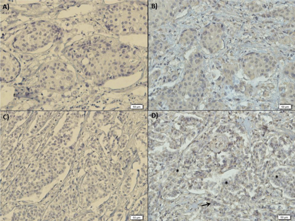
| Figure 3: Immunohistochemical assay with the polyclonal antibody anti-ADAM33. Ductal samples were composed of cells with marked atypia, hyperchromatic nuclei and coarse chromatin, ample cytoplasm and indistinct boundaries. A) IDC negative control, B) IDC anti–ADAM33: cytoplasmic and weak membrane signal positivity for antibody evidenced by the brown color of these structures. For lobular samples we observed atypical neoplastic cells arranged in solid aggregates or in a single row, and a little desmoplastic matrix, interspersed in the neoplastic lobules. C) ILC negative control, D) ILC anti–ADAM33: poor response to polyclonal anti-ADAM33 antibody showing the region with invasive lobular cancer cells in a single row (arrow), * Invasive lobular cancer cells in aggregates, 200 X. |