|
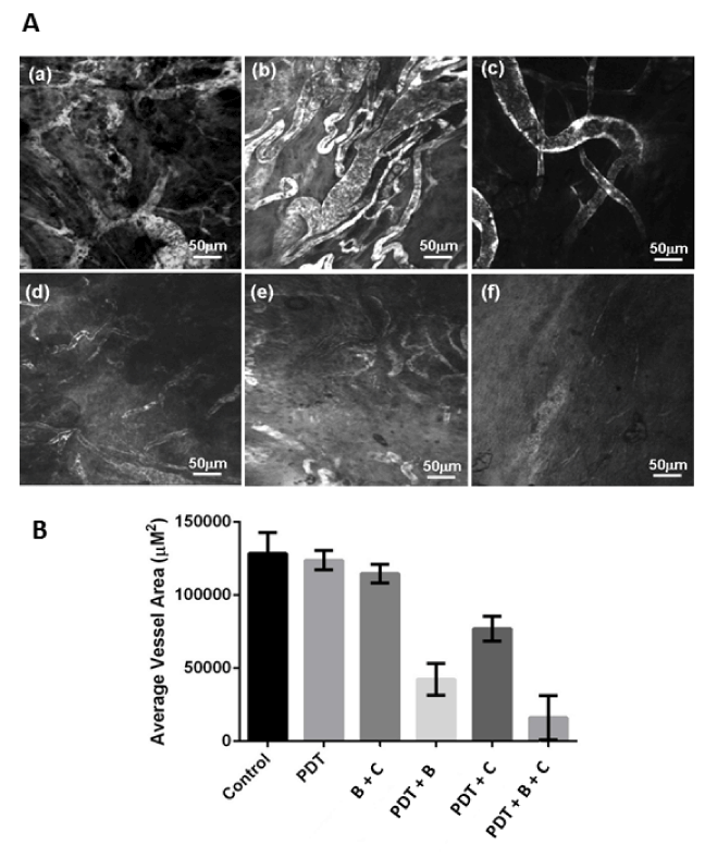
| Figure 5: (A): Tumor blood vessels visualized approximately 30 min after intravenous administration of FITC-dextran in (a) control, (b) PDT only, (c) Bevacizumab + cetuximab, (d) PDT + bevacizumab, (e) PDT + cetuximab and (f) PDT + bevacizumab + cetuximab treated models using confocal endomicroscopy. Magnification X200, scale bar = 50 μm. (B) Average vessel area for each animal group was calculated from images captured from three locations with high vessel density and that are 20 and 40 μm below the surface of the tumor. |