 |
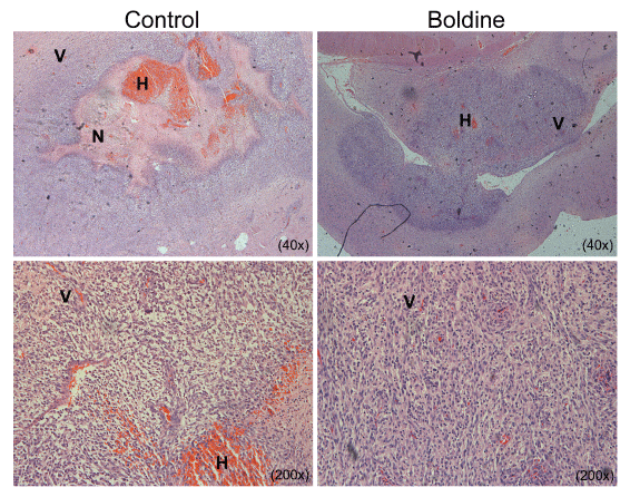
| Figure 5: Histological analysis of implanted gliomas. The sections of implanted rat glioma were stained with hematoxylin and eosin (H&E), as described in Section 2. Representative pictures of histological characteristics seen in rats of control group and treated group. Legends: (N) necrosis, (V) microvascular proliferation, hemorrhages (H), Magnification 40x, 200x. |