|
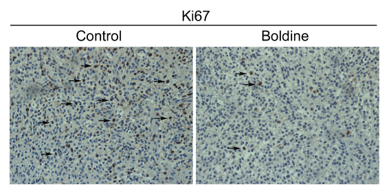
| Figure 6: Immunohistochemical staining of Ki67 in implanted gliomas. Glioma cell proliferation was assessed by immunostaining for Ki67 positive glioma cell nuclei (arrows). Representative pictures of immunohistochemical analysis in control group and in boldine-treated group. Magnification 400x. |