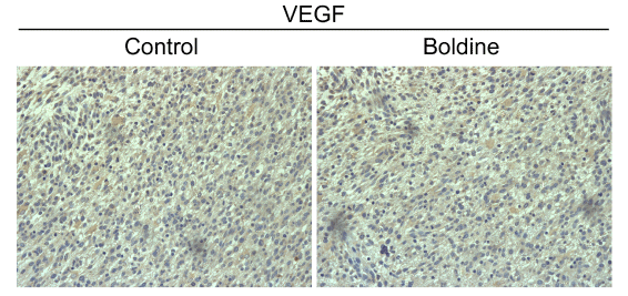
Figure 7:
Immunohistochemical staining of VEGF in implanted gliomas. Glioma cell angiogenesis was assessed by VEGF staining. Representative pictures of immunohistochemical analysis in control group and in boldine-treated group. Magnification 400x.