 Lane 1: TLR-3; Lane 2: TLR-4 and Lane 3: TLR-9;
Lane 1: TLR-2; Lane 2: TLR-3; Lane 3: TLR-4; Lane 4: TLR-9. In the case of OC-3-VGH ovarian cancer cells, the expression of TLR-2 gene is too low to be quantitated.
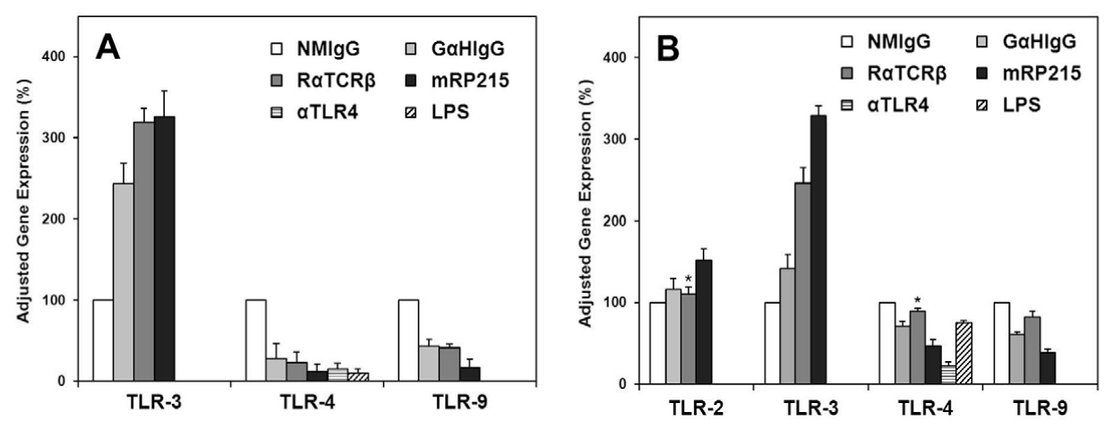
All data presented are statistically significant at P<0.01 except those labeled with * which are not statistically different from the negative control.
Lane 1: TLR-3; Lane 2: TLR-4 and Lane 3: TLR-9;
Lane 1: TLR-2; Lane 2: TLR-3; Lane 3: TLR-4; Lane 4: TLR-9. In the case of OC-3-VGH ovarian cancer cells, the expression of TLR-2 gene is too low to be quantitated.
All data presented are statistically significant at P<0.01 except those labeled with * which are not statistically different from the negative control. ), anti-TLR-4
(αTLR4) (
), anti-TLR-4
(αTLR4) ( ) and LPS (
 ) with normal mouse IgG (NMIgG) was adjusted to 100% in all cases.