 |
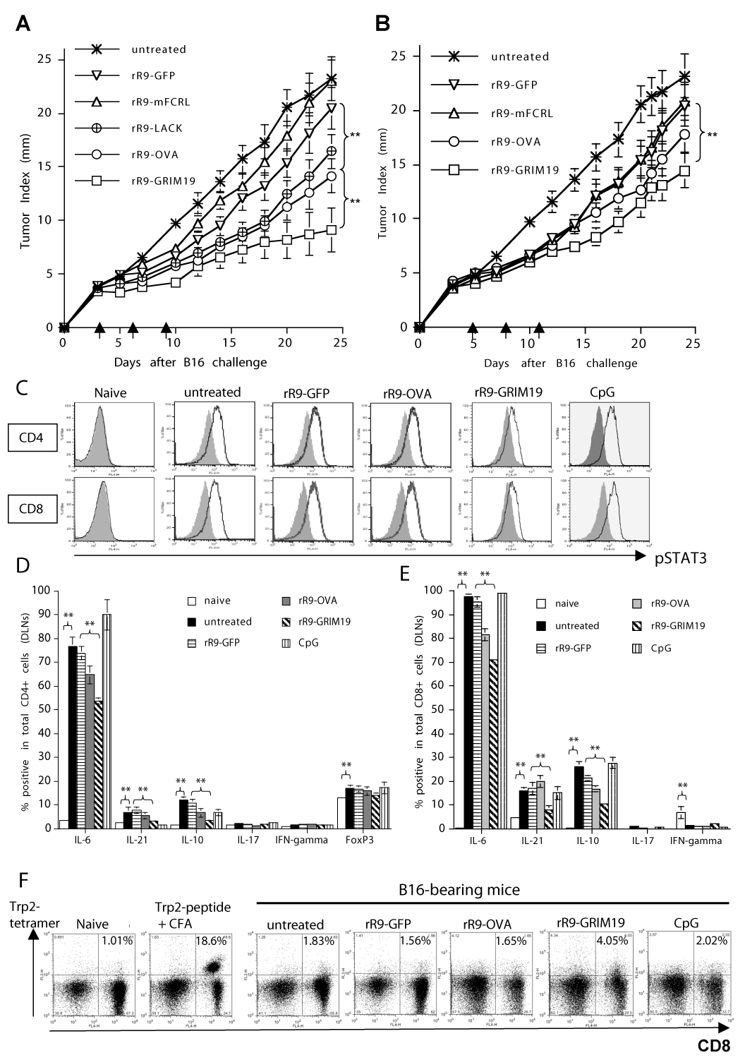
| Figure 2: Relationship between antitumor effects and pSTAT3 expression/ cytokine-profile in T cells after treatment with rR9-fusion proteins in B16 melanoma–bearing mice. (A, B): Anti-B16 tumor effects by intratumoral injections of rR9-fusion proteins on days 3, 6, 9 (A) or on days 5, 8, 12 (B). rR9-OVA was severely diminished when immunizations were started on or after day 5 (B), but not on or after day 3 (A). (** p < 0.01). Data are representative of three individual experiments. (C): Expression of pSTAT3 in CD4+/CD8+ T cells from DLNs following intratumoral injections of rR9-fusion proteins or CpG on days 5, 8 and 12; data were collected on day 16. Gray lines, isotype IgG. Data are representative of three individual experiments. (D): A summary of frequencies of IL-6/IL-21/IL-10/IL-17/IFN-γ cytokine-producing or FoxP3+ CD4+ cells (% of positive cells by intracellular staining with specific mAbs) following intratumoral injections of rR9-proteins in DLNs of B16- bearing mice on day 13. Data are representative of three individual experiments. (** p < 0.01). (E): A summary of frequencies of IL-6/IL-21/IL-10/IL-17/IFN-γ cytokineproducing CD8+ cells following intratumoral injections of rR9-proteins in DLNs of B16-bearing mice on day 13. Data are representative of three individual experiments. (** p < 0.01). (F): Frequencies of CD8+ Trp2-tetramer+ cells in DLNs of B16-bearing mice following intratumoral injections of rR9-proteins/CpG on day 13. Data are representative of two individual experiments. |