|
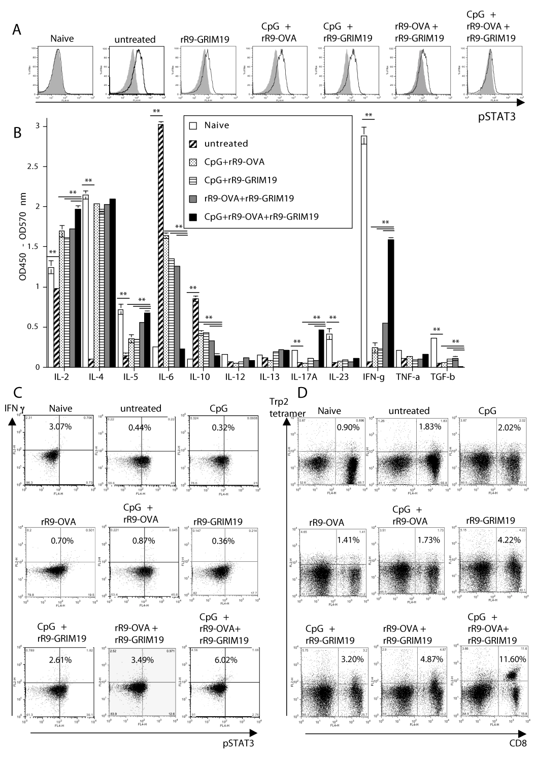
| Figure 5: Analyses for pSTAT3 expression and cytokine-profile in CD8+ T cells from COG-treated B16 melanoma-bearing mice. (A): Expression of pSTAT3 in CD8+ T cells from DLNs following intratumoral injections of CpG, rR9-OVA and/or rR9-GRIM19 on days 5, 8 and 12; data were collected on day 16. Gray lines, isotype IgG. Data are representative of three individual experiments. (B): Screening of cytokine profiles in CD8+ T cells from DLNs of B16-bearing mice following intratumoral injections of CpG, rR9-OVA and/or rR9-GRIM19. Purified CD8+ cells that were harvested on day 13 from DLNs were restimulated with PMA and ionomycin for 48 h in vitro. Then, cell supernatants were analyzed with multiple cytokine ELISA analyses. Data are representative of two individual experiments. (** p < 0.01). (C): Expression of pSTAT3 and IFN-g in CD8+ T cells from DLNs following i.t. injections of CpG, rR9-OVA and/or rR9-GRIM19. Cells that were harvested on day 13 were restimulated with PMA and ionomycin for 48 h in vitro, and intracellular staining was performed with mAbs. Data are representative of three individual experiments. (D): Frequencies of CD8+ Trp2180-188-tetramer+ cells following intratumoral injections of CpG, rR9-OVA, and/or rR9-GRIM19 in DLNs of B16 melanoma-bearing mice on day 13. Data are representative of three individual experiments. |