 |
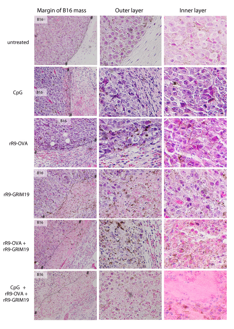
| Figure 6: Histopathological analyses of B16 melanoma-bearing area after treatments. Skin biopsies were performed from B16 tumor-bearing skin on day 13 after treatments with CpG, rR9-OVA, rR9-GRIM19, rR9-OVA+rR9-GRIM19, or COG on days 5, 8, and 12. Vertical skin sections of the samples were stained with H & E (original magnification; ×40 and ×100). The margin, outer layer and inner layers of B16 tumor mass are shown. Dotted lines with # represent the margin of B16 mass. Data are representative of two individual experiments. |