|
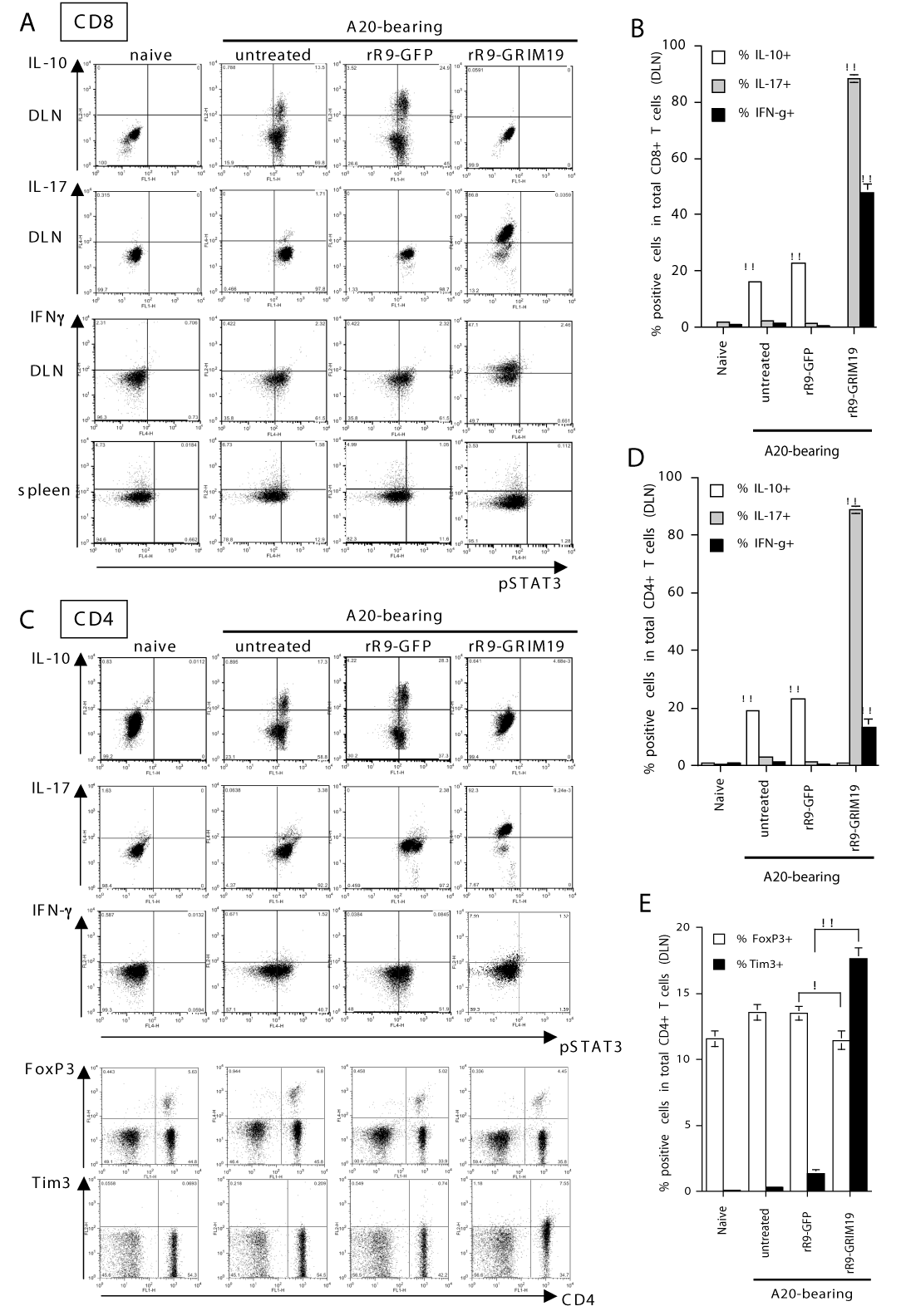
| Figure 5: Analyses of CD8+ or CD4+ T cell subsets after intratumoral injections of rR9-GRIM19. A20-bearingmice were treated with rR9-GFP or rR9-GRIM19 injected i.t. daily on days 9, 10, 11. DLNs or spleens were collected from untreated, rR9-GFP-treated, or rR9-GRIM19-treated A20 tumor-bearing mice. (A): Expression of pSTAT3 and production of IL-10/ IL-17/ IFN- γ in CD8+ T cells from DLNs and spleen following i.t. injections of rR9-fusion proteins. Cells that were harvested on day 13 were restimulated with PMA and ionomycin for 48 h in vitro, and intracellular staining was performed using mAbs. Data are representative of threeindividual experiments. (B): A summary of the average percentages of CD8+T cells from DLNs (on day 13) that produced IL-10-, IL-17-, or IFN- γ is shown in Figure 5A. (**; p <0.01) (C): IL-10- /IL-17- /IFN-γ-producing, FoxP3-positive, or Tim3-positive purified CD4+ T cells in DLNs after intratumoral injections of rR9-fusion proteins (data were collected on day 13). The harvested cells were restimulated in vitro with PMA and ionomycin, and intracellular staining was performed with mAbs. Data are representative of three individual experiments. Data from only one representative experiment is shown. (D): Summary of the average percentages of IL-10-/ IL-17-/ IFN- γ -producing CD4+ cells in DLNs (on day 13). (**; p < 0.01) (E): A summary of the average percentages of FoxP3+ CD4+ or Tim3+ CD4+ T cells in DLNs (on day 13). (*; p < 0.05, **; p < 0.01) |