|
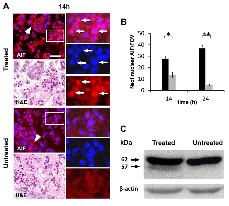
| Figure 7: Activation of apoptosis inducing factor (AIF) upon mEHT treatment of HT29 xenografts. A) Identical tissue sections consecutively stained for AIF using immunofluorescence (Alexa 564, red) then with H&E. Many tumor cells show nuclear translocation of AIF in the treated but not in the untreated tumors. Insets show single channel views at higher power of representative areas within rectangles. Arrows highlight identical cells with nuclear AIF staining. Arrowheads points to tumor nests of granular mitochondrial AIF staining characteristic of intact tumor cells. Cell nuclei are stained using DAPI (blue). Bar indicates 50 μm in the left and 25 μm in the right column. B) Graph showing the significantly elevated mean number of nuclear AIF positive cells in the treated (black columns) compared to the untreated (grey columns) tumors (*p<0.05; **p<0.001). C) In western immunoblots the 57kDa cleaved AIF protein is detected only in the treated tumors at 14h post-treatment besides the mature 62kDa protein. |