 |
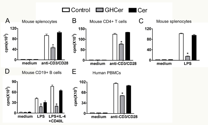
| Figure 2: Inhibition of proliferation of immune cell by GHCer. Indicated immune cells were incubated with GHCer, ceramide or medium control for 24 hrs and then activated by anti-mouse CD3 and CD28 mAbs (A and B), LPS (C), LPS in the presence of IL4 and mouse CD40 ligand (D), or anti-human CD3 and CD28 mAbs (E). Cell proliferation was determined by [3H]-thymidine incorporation assay. The data are presented as mean ± SD of triplicate. *, p<0.05, compared with control. |