|
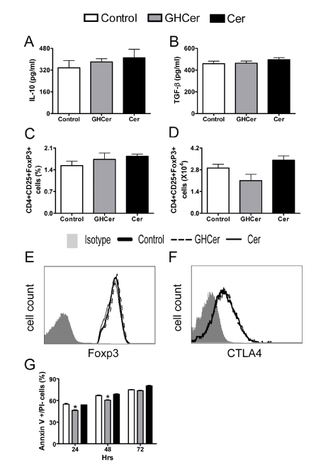
| Figure 4: GHCer could neither expand Treg population nor induce apoptosis of splenocytes. Mouse splenocytes were incubated with GHCer, ceramide or medium control for 24 hrs, before activation by anti-mouse CD3/CD28 for 3 days. Supernatants were collected for determination of IL-10 (A) and TGF-β (B) by ELISA assay. Cells were stained with antibodies against CD4, CD25, Foxp3 and CTLA4 at day2 and the percentage (C), absolute number (D), expression of Foxp3 (E) and CTLA4 (F) of Treg cells were determined. Splenocytes were harvested at the indicated time points for determination of apoptotic cells by staining with annexin V and PI, followed by flow cytometric analysis. The percentage of apoptotic (annexin V+/PI-) cells was shown (G) and data are presented as mean ± SD of triplicate. *, p<0.05, compared with control. |