|
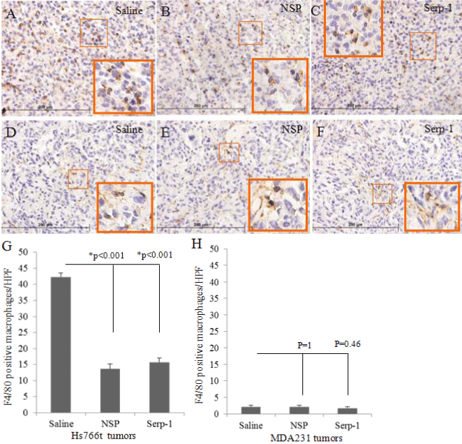
| Figure 6: Serpin treatment decreased macrophage infiltration in pancreatic cancer xenografts. Macrophages in the tumor tissue were stained with F4/80 antibody. Positive cells in five randomly selected high power fields (HPFs) were counted for each tumor section. The averages for positive cell counts from each treatment group were compared by ANOVA. (A-C) Representative tumor sections immunostained for macrophages (brown stain) in pancreatic cancer tissue are provided from saline (A), NSP (B) and Serp-1 (C) treatment groups. (D-F) Positively stained macrophage cells in breast cancer tissue from saline (D), NSP (E), and Serp-1 (F) treatment groups are also shown. (G) Positively stained macrophages in pancreatic cancer tissues were significantly decreased after serpin treatment (P<0.001, n=40 HPFs total). (H) However, serpin treatment did not significantly change macrophage number in breast cancer xenografts (P>0.4, n=35 HPFs total). Magnification 400X for histology sections and 1000X for higher power insets for each stained section * indicates significance of P ≤ 0.05. |