|
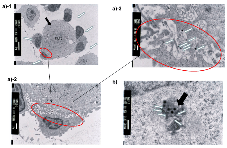
| Figure 7: To determine cellular localization of internalized Ad5/F35 particles, PC3 cells were evaluated using electron microscopy. Ad vector particles were observed within endocytic vesicles (a) (white arrow indicates γδ T cells, black arrow indicates PC3 cells). Ad vectors were observed between adjacent γδ T cells and PC3 cells (b) (white arrows indicate Ad vectors). Multivesicular bodies (MVBs) were observed within the cytoplasm of PC3 cells (black arrow), and Ads were recognized within MVBs (white arrow) (c). |