 |
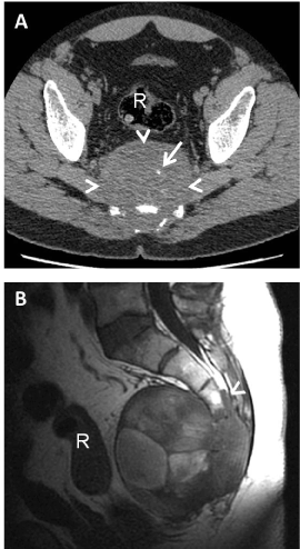
| Figure 1: The primary chordoma. (A) Axial non-enhanced CT image. There is a mass (arrowheads) destroying the sacrum and growing towards presacral region. The mass contains small calcifications (arrow). The rectum (R) is located anteriorly. (B) MR Sagital SSFSE T1 weighted image. The MR shows a heterogeneous mass which invades the sacral canal (arrowhead). The mass has high signal intensity contents that probably represent hemorrhagic components. Rectum (R). |