|
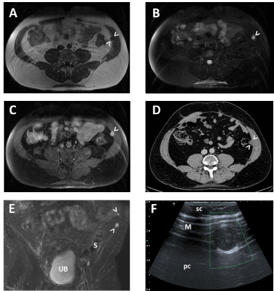
| Figure 2: Metastases of chordoma in the abdominal wall. (A) MR Axial SSFSE T1 weighted image. There is a nodule in contact with the internal side of the left transversalis muscle of the abdominal wall (arrowheads). The nodule is hypointense to the muscle and is barely distinguished from the transverse abdominus. (B) MR Axial SSFSE T2 weighted image with fat saturation. The nodule is markedly hyperintense (arrowhead). (C) MR Axial SSFSE T1 weighted image with fat saturation and intravenous gadolinium contrast injection. The nodule shows a peripheral rim enhancement (arrowhead). (D) Axial enhanced CT image. Nodule in contact with the internal side of the left transversalis muscle (arrowheads). (E) MR Coronal SSFSE T2 weighted image with fat saturation. The two abdominal wall metastases are pointed with arrowheads. Urinary bladder (UB) and sigmoid colon (S). (F) Doppler ultrasound image of one of the abdominal wall nodules, obtained with a linear transducer. There is no evidence of significant flow inside the nodule. Abdominal muscle wall (M), subcutaneous tissue (sc) and peritoneal cavity (pc). |