 |
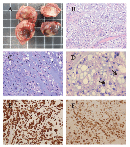
| Figure 3: Histopathological analyses of the surgical specimen. Metastatases of chordoma in the abdominal wall (A) Gross image of chordoma showing a lobulated and well-defined tumor. (B) Hematoxylin and eosin-stained sections of tumor with a lobular growth pattern (X20). (C) Abundant basophilic to metachromatic mucinous or myxoid stroma comprises the majority of the tumor volume. The cells of chordoma are round to ovoid with pale eosinophilic cytoplasm growing in strands, cords, and clusters (X200) (D) Physaliphorous cells are large cells with multiple vacuoles in the cytoplasm (X400). (E) In this case the neoplastic cells are immunoreactive strongly with CKAE1/AE3, and (F) protein S-100 in a weak and patched (X200). |