|
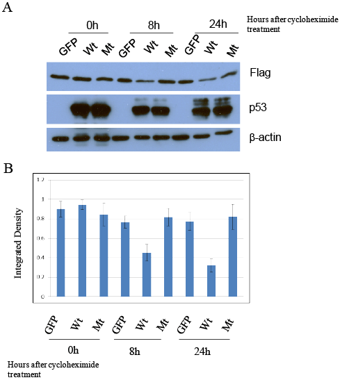
| Figure 3: Wild type p53 but not mutant p53 (R175H) suppressed exogenous 14-3-3γ protein. (A) H358 cells that stably expressed Flag-14- 3-3γ protein were infected by Ad-GFP, Ad-p53 (wt) and Ad-p53 (mt). 24 h after infection, the cells were treated with 20 µg/ml cycloheximide for 0 h, 8 h and 24 h and then harvested for detecting Flag-14-3-3γ protein level by immunoblotting. Both wild type and mutant p53 protein were also determined. β-actin was used as a loading control. The experiment was repeated three times and a representative data was shown. (B) The expression of Flag-14- 3-3γ protein seen in panel A was quantitated on a Strantagene Eagle Eye II, normalized against β-actin and graphed. The graph represents the mean number of the integrated density ± S.D. (n=3). |