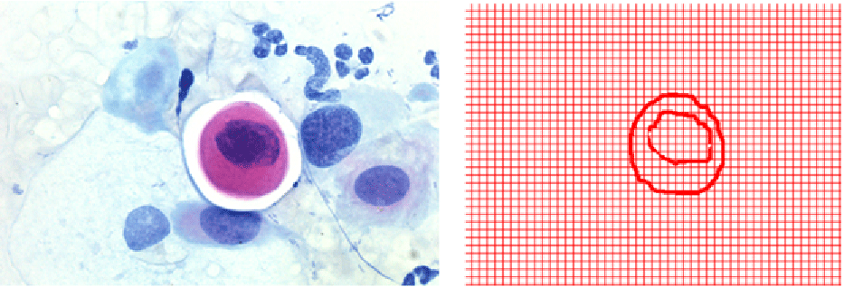
Figure 1:
Original image (left), and Image obtained by the software (right), of the nucleus and cytoplasm frontiers of an H-SIL cell. The red lines correspond to the grid of 2 pixel´s side.