|
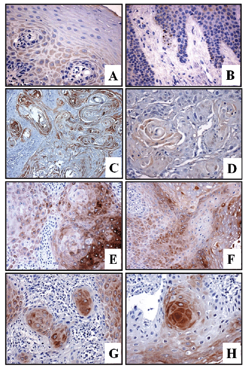
| Figure 1: Examples of immune histochemical staining for CD133 in normal oral epithelium (A, B) and OSCC (C-H). (A) Normal squamous epithelium displaying a diffuse basal and para-basal immunopositivity (x200). (B) Normal squamous epithelium with immunopositivity localized to the basal layer (x150). (C) A well-differentiated OSCC with a diffuse immunopositivity (x100). (D) A moderately differentiated OSCC with a focal immunopositivity (x200).(E-H) Different OSCC samples with an intense immunopositivity for CD133, localized primarily in epithelial nests infiltrating surrounding stroma (x200). |