 |
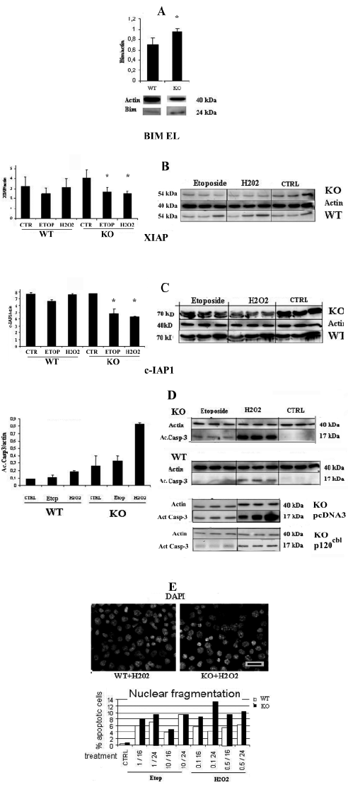
| Figure 2: c-Cbl protects primary mouse embryonic fibroblasts (MEFs) predominantly against oxidative stress. One representative in vitro experiment is reported. The triplicate mean of protein expression level +/- s.e.m. is given. Etoposide were done at different concentrations. The optimal results were obtained with the indicated concentration. a: A) Bim EL is significantly higher in c-Cbl KO (p = 0.032) compared to WT cells. B and C) The inhibitors of apoptosis XIAP and c-IAP1 expression in MEFs untreated (CTRL) or treated by 0.1 mM hydrogen peroxide (H2O2) for 24 hours were significantly lower than control in KO-MEFs for XIAP (H2O2 p = 0.0028; Etoposide p = 0,038) and for c-IAP1 (H2O2 p = 0.0016; Etoposide p = 0.023). b. D) Activated caspase-3 is strongly activated in KO-MEFs compared to WT (p < 0.001) only in the presence of 0.1 mM of H2O2. Etoposide gave a much lower activation of activated caspase-3. The two right upper panels show the results of KO and WT MEFs, the two panels below, those of MEFs transfected with the vector alone (pcDNA3) and the c-Cbl expression vector. These different experiments gave very similar results. E) Nuclear fragmentation of the KO and WT MEFs revealed by DAPI staining experiments at diverse indicated concentrations (mM for etoposide, mM for H2O2) and after 16 or 24 hours of indicated treatment. The most effective concentrations for nuclear fragmentation were 1mM etoposide and 0.1 mM H2O2 after 24 hours of treatment. On the upper panel, DAPI staining of MEF nuclei. Magnification bar = 20mm. |