|
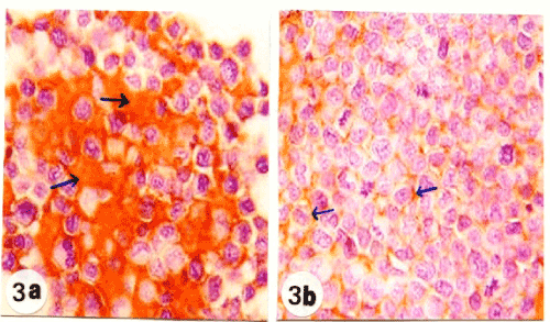
| Figure 3: Photomicrographs of EAC-sections stained immunohistochemically to show the higher concentration of Bcl-2 (arrow; brownish yellow color) in the cytoplasm of EAC-cells in control mice (figure 3a; x100) as compared with mice treated with ulvan polysaccharide (figure 3b; x100). |