|
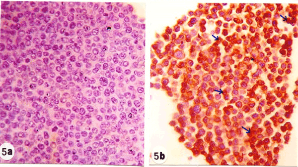
| Figure 5: Photomicrographs of immunohistochemically stained EAC-sections, showing the higher amount of TdT (arrow; brownish yellow color) in the nuclei of EAC-cells in mice treated with ulvan polysaccharide (figure 5b; x100) than in control mice (figure 5a; x100). |