 |
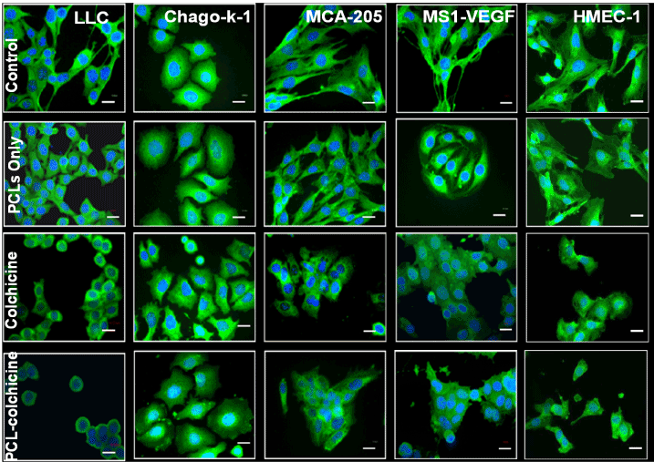
| Figure 2: Fluorescence images of endothelial cell, HMEC-1, 24 hrs following exposure to colchicine (free drug) or various PCLs (with colchcine). The images show qualitative effect of different cationic lipids of PCLs on cytoskeleton (green) and the nucleus (blue) cell areas. Images were captured using 40X magnification. Magnification bar = 20 µm. |