|
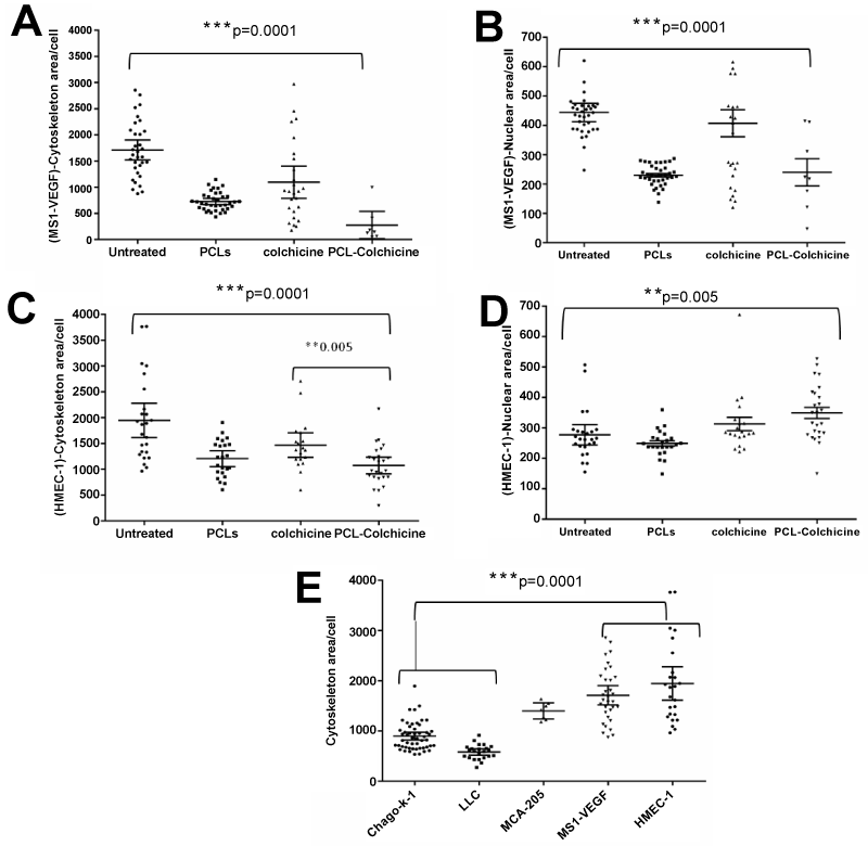
| Figure 4: Quantitative analysis of cytoskeleton area per cell (A, C) and nuclear area per cell (B, D) of the endothelial cell lines, MS1-VEGF (A, B) and HMEC-1 (C, D) are depicted in the form of scatter plots.). E) Quantitative comparison of cytoskeleton area per cell of all the cell lines screened in this study. Each dot on the plot reflects a single field in the cell sample. The statistics were performed using one way ANOVA and the error bars represent the 95% CI. Bonferonni post hoc tests were used to compare all the pairs of columns to determine the p value. ** p < 0.01. |