 |
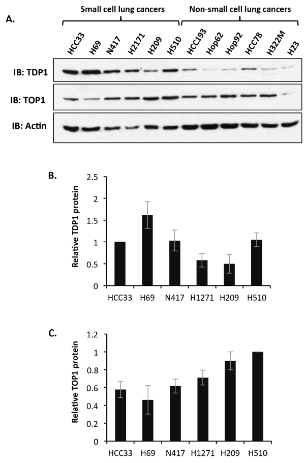
| Figure 1: Small cell lung cancer cell lines display remarkable variation in both TDP1 and TOP1 protein level. (A) Whole cell extracts (40 μg) from the indicated small cell lung cancer cell lines and other non-small cell lung cancer cells were separated by 10 % SDS-PAGE and immunoblotted using antibodies against TDP1, TOP1 and actin. A quantitation of relative TDP1 (B) and TOP1 (C) protein level is depicted. Data are the average of 3 independent experiments ± STD normalised to HCC33 for TDP1 and H510 for TOP1. |