|
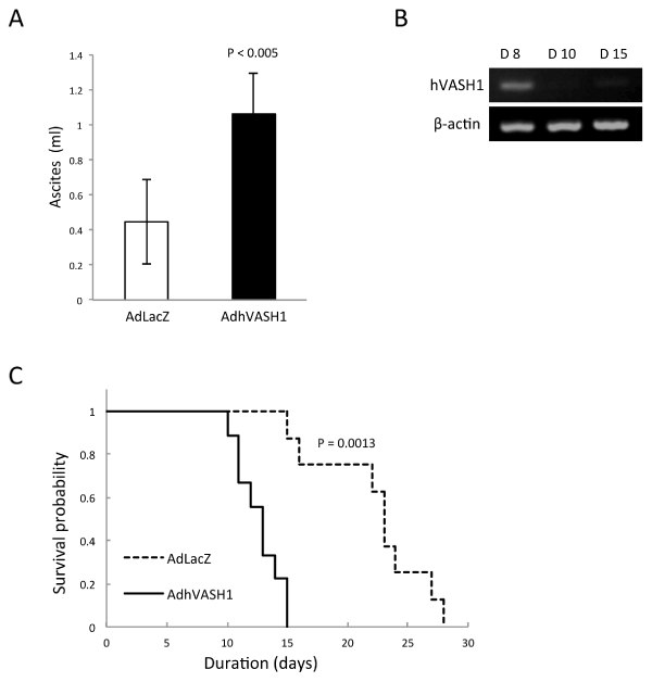
| Figure 3: HM-1/hVASH1 tumors resulted in a worse outcome when inoculated into the peritoneal cavity. A: HM-1/LacZ or HM-1/hVASH1 cells were inoculated into the peritoneal cavity of mice (N=4). Eleven days after the inoculation, the ascites fluid was collected and its volume measured. Means and SDs are shown. B: The time course of expression of hVASH1 in the inoculated HM-1/hVASH1 cells was examined by RT-PCR. C: Survival probability was compared between mice with HM-1/ LacZ (N=8) and mice with HM-1/hVASH1 (N=8). |