 |
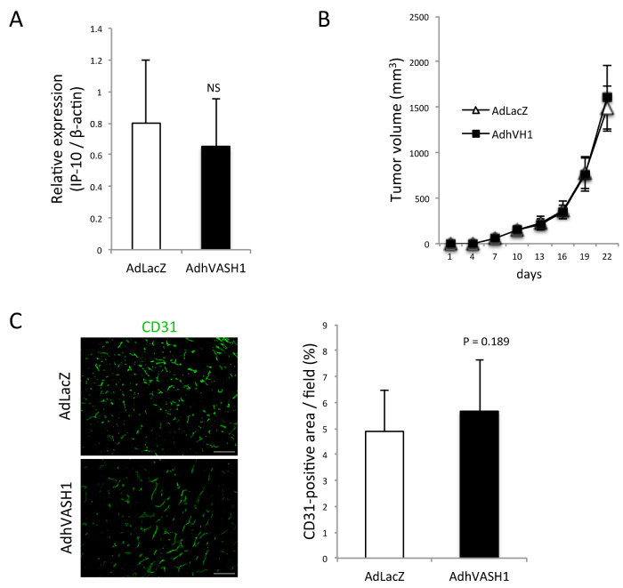
| Figure 6: Tumor growth and tumor angiogenesis were unchanged when HM-1/hVASH1 cells were subcutaneously inoculated in SCID mice. A: SCID mice were subcutaneously inoculated with HM-1/LacZ (N=5) or HM-1/hVASH1 (N=5) cells. The expression of IP-10 in the tumors was compared by quantitative RT-PCR. Means and SDs are shown. B: SCID mice were subcutaneously inoculated with HM-1/LacZ (N=5) or HM-1/hVASH1 (N=5) cells, after which the tumor size was measured. C: The tumor microvessel density was assessed on day 28 post inoculation by immunostaining with anti-CD31 Ab. Scale bar=200 μm. H: The CD31-positive vascular area was quantified and compared between HM-1/LacZ (N=4) and HM-1/hVASH1 (N=4) tumors. Means and SDs are shown. |