|
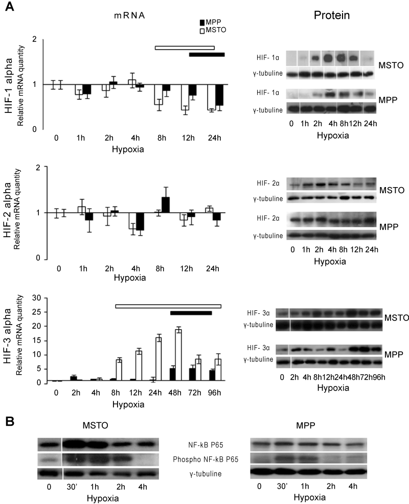
| Figure 1: HIF-1, HIF-2, HIF-3 alpha and NF-κB expression profiles in MSTO and MPP cell lines in hypoxic conditions. (A) On the left side, the mRNA expression levels of HIF-1, HIF-2, and HIF-3 alpha at different times of hypoxia were evaluated through RT PCR. Values are expressed as fold induction with respect to normoxic control (set at 1) and represent the mean ± SE of three different experiments. 18S rRNA was used as houseκeeping gene. Horizontal bars at the top of each graphic indicate significant values (P≤0.05). On the right side, the nuclear levels of HIF-1, HIF-2, and HIF-3 alpha proteins at different times of hypoxia were analyzed by Western Blot. γ-tubuline was used as loading control. A representative experiment for each gene is shown. (B) The nuclear levels of NF-κB P65 and phospho NF-κB P65 (Ser276) at different times of hypoxia were analyzed by Western Blot in MSTO and MPP. γ-tubuline was used as loading control. A representative experiment is shown for both NF-κB P65 and phospho NF-κB P65 (Ser276). |