|
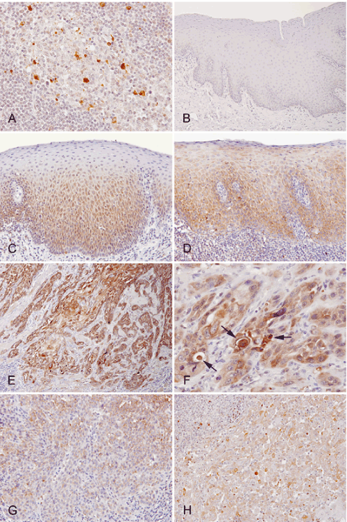
| Figure 1: Immunohistochemical localization of cleaved caspase-3 in non-neoplastic epithelia, epithelial dysplasia, carcinoma in situ (CIS), invasive squamous cell carcinoma (SCC), and metastatic SCC in samples of lymph nodes. The representative staining pattern of cleaved caspase-3 in the germinal center of non-metastatic lymph node (A), non-neoplastic epithelium (B), epithelial dysplasia (C), CIS (D), invasive carcinoma (E-G), metastatic carcinoma samples (H); (A–H) immunoperoxidase stain for cleaved caspase-3. Specific positive staining for cleaved caspase-3 was confirmed in apoptotic lymphocytes in the germinal centers of lymph nodes (A); however, no obvious positive staining in non-neoplastic squamous epithelia was observed (B). In epithelial dysplasia, cleaved caspase-3 was localized in the proliferative foci of parabasal cells in the lower half of the epithelia (C). In CIS, positive staining was observed in almost the entire layer of the epithelia except for the superficial keratinized cells (D). In invasive carcinoma, extensive positive staining was observed in carcinoma cells (E). The cytoplasm of carcinoma cells lacking apoptotic figures was diffusely positive for cleaved caspase-3 in addition to apoptotic carcinoma cells showing a rounding of their cytoplasm and nuclear condensation (F). Arrows indicate apoptotic carcinoma cells. Staining intensities varied from case to case, and in particular, less keratinized carcinoma cells displayed weak staining (G). Metastatic carcinoma cells in the lymph nodes were also positive for cleaved caspase-3 (H). |