|
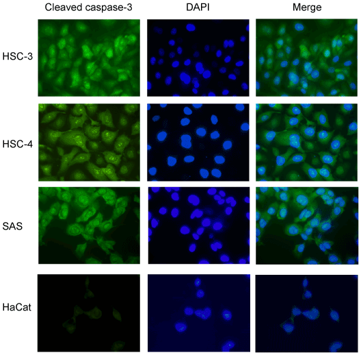
| Figure 2: Subcellular localization of cleaved, active caspase-3 in oral squamous cell carcinoma (OSCC) cells. HSC-3, HSC-4, SAS, and HaCat cells were cultured on glass chamber slides overnight at 37°C. They were then analyzed using CellEvent® Caspase-3/7, a green detection cellpermeable reagent for the intracellular distribution of cleaved caspase-3, under a fluorescence microscope. Green indicates cleaved caspase-3 and blue indicates nuclei. |