|
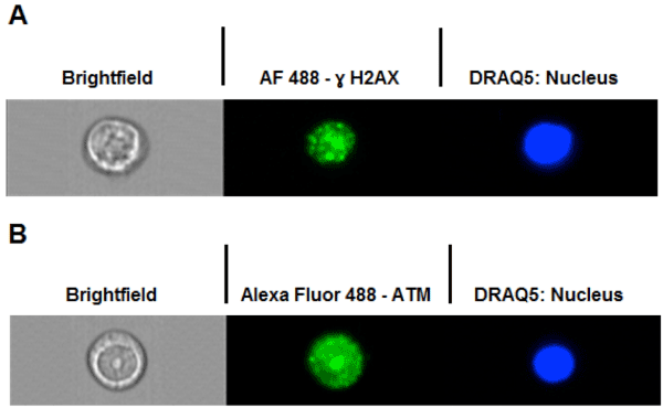
| Figure 5: A and B Show representative images of imaging flow cytometry of γ-H2AX foci staining (A) and total ATM staining (B). Three channels are presented whereby channel one represents the brightfield image; channel two the immune-labelled γ-H2AX foci or ATM protein and channel 5 shows the DRAQ5 staining of the nucleus within the cells. |