|
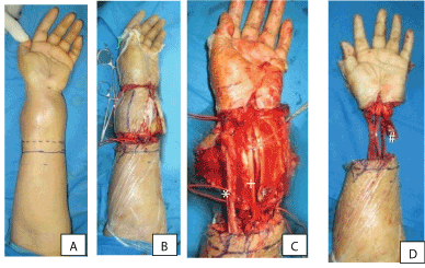
| Figure 7: Intraoperative pictures. (A) The circumferencial design; (B) The skin incision that made accordingly; Identification of the structures of the foream. (C) Preservation of the neurovascular bundle of the distal forearm. (asterisk; superficial radial nerve and radial artery, plus sign; median nerve, pound sign; ulnar nerve and ulnar artery); (D) After tumoural resection (osteotomized the radius and ulna), cut the tendons and performed disarticulation of the left wrist joint. |