|
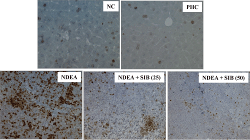
| Figure 10: Cell proliferation was assessed by Ki67 immunolabeling in the liver Normal, NDEA treated and SIB treated animals. NDEA-Treated animals at the end of 8th week of induction revealed a large number of Ki67- labeled hepatocytes; In normal control (NC) and PH-control (PHC) group of animals showed the vast majority of hepatocytes Ki67-negative; 2 weeks of treatment with SIB reduced the Ki67 positive hepatocytes in liver in a dose dependent manner. Original magnification: ×40. Hepatocytes proliferation was measured as the proportion of Ki67-positive hepatocytes. |