 |
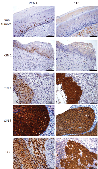
| Figure 1: Representative image of immunohistochemical analysis of p16INK4a (p16) and proliferating cell nuclear antigen (PCNA) expression in cervical tissue (x200). Non-tumoral and CIN 1 show nuclear PCNA staining with absence of p16 signal. Varying degree of staining intensity was observed in cervical intraepithelial neoplasia (CIN 1/2). Both CIN 3 and invasive squamous cell carcinoma tissues displayed diffuse and strong staining for all markers. |