|
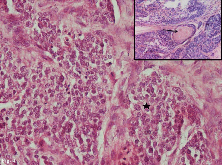
| Figure 4: Histosections show tumor cells with round bland nuclei, inconspicuous nucleoli and moderate pale eosinophilic to clear cytoplasm and distinct cell outline (star). The cells were arranged in lobules and ducts some of which contained eosinophilic secretion (Inset, arrow) (H and E, x400). |