|
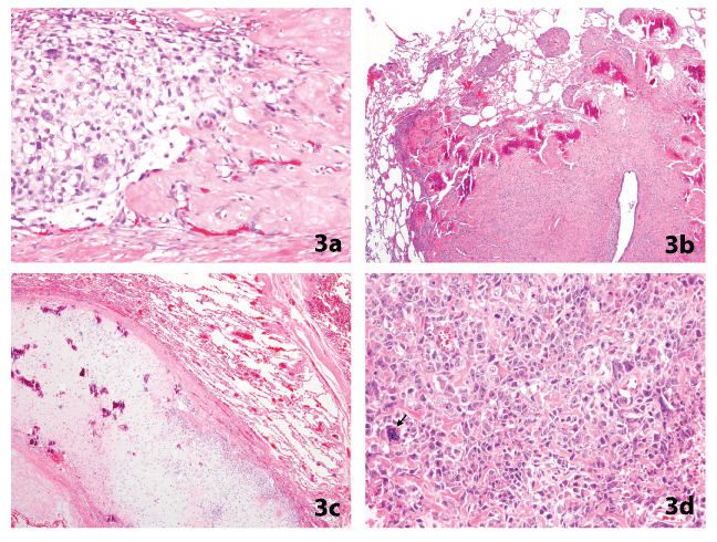
| Figure 3a,b,c,d: Osteosarcoma in the lung. Photomicrographs of hematoxylin and eosin stained slides at medium magnification (objective lens x10) shows malignant cells forming osteoid (3a) with an osteoblastic matrix in Figure 3b and a chondroblastic matrix in Figure 3c. High magnification (objective lens x20) in Figure 3d shows the presence of malignant hyperchromatic cells with marked cellular and nuclear atypia with abnormal mitosis (▼). |