|
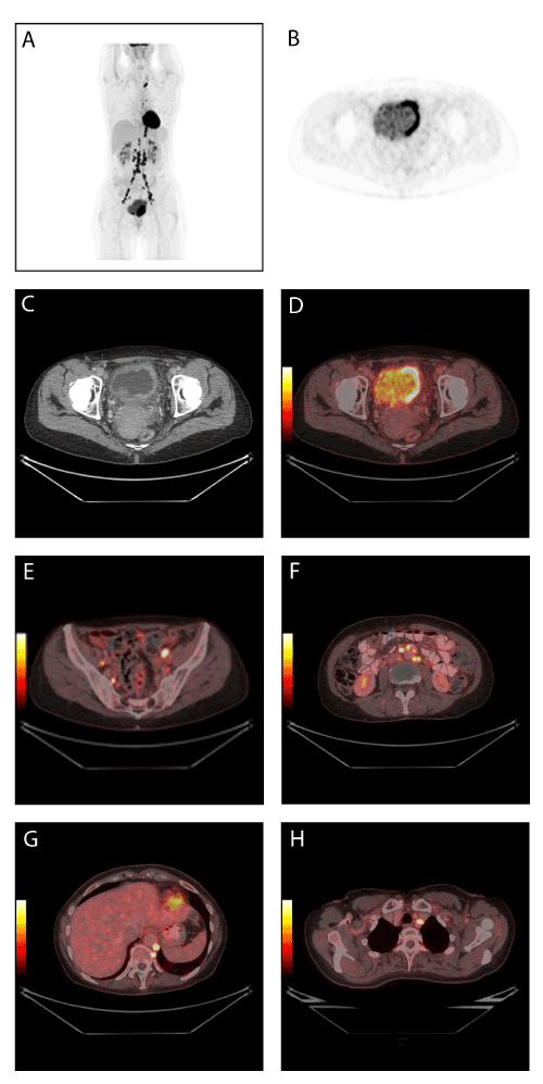
| Figure 2: A 56-year-old patient with newly diagnosed muscle invasive bladder cancer underwent 18F-FDG PET/CT for staging. The PET image shows the tumour in the bladder as well as multiple lymph node metastases (2A). The tumour can be seen in the left side of the urinary bladder on axial PET image (2B), CT image (2C), and fused PET/CT image (2D). Multiple metastases are seen in lymph nodes located in both sides of the pelvic area (2E), in retropetoneal lymph nodes (2F), in retrocrural lymph nodes and in one lymph node on the left side of trachea (2G). |