|
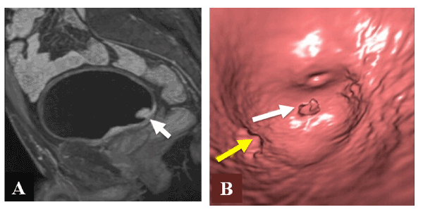
| Figure 5: 62-year-old patient with hematuria. Sagittal contrast enhanced T1W MR image (A) demonstrated an enhancing papillary lesion consistent with bladder cancer (white arrow), virtual MR cystoscopy MR image (B) confirms the presence of a papillary bladder cancer lesion (white arrow), near the orifice of the left ureter (yellow arrow) (B). (Courtesy of Dr. Evrim B. Turkbey from Radiology and Imaging Sciences Department, Clinical Center NIH, Bethesda, MD, USA). |