|
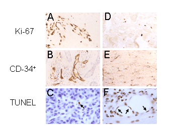
| Figure 2:Numerous Ki-67 immunostained cells (brown) are selectively detected in the pre-treated specimen (Panel A vs. Panel D). CD34+ immunostaining (brown) reveals irregularly dilated vessels in pre-treated tumors (Panel B), which are absent in post-treated tumors (Panel E). A clear increase in the number of TUNEL-positive cells (brown) is observed following treatment in the hypodermis (Panel C vs. Panel F). Endothelial apoptotic nuclei in panel N are indicated by arrows. Panels C and F were counterstained with hematoxylin. Original magnification (x200). |