|
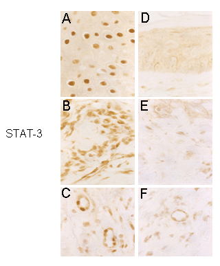
| Figure 3: Immunohistochemical staining of Tyr705-phosphorylated STAT-3 in biopsies obtained before (panels A-C) and after the three month treatment (panels D-F). Sections correspond to the superficial (A and D) medium (B and E) and deep dermis (C and F), respectively. In panel C two neovessels in the dermis show STAT-3 immunoreactivity in endothelial cell nuclei. All the shown pictures used to exemplify the effect of the treatment belong to the same patient.; Original magnification (x 400). |