|
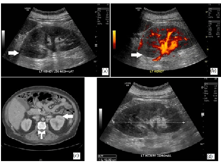
| Figure 1: Figure shows (a) Acute lobar nephronia (arrow) in the upper pole of left kidney on ultrasound, (b) Power Doppler evaluation revealing marginal reduction in perfusion to the upper pole of the left kidney, (c) Focal lobar nephronia on CT abdomen, and (d) Resolution of nephronia in the left kidney six weeks after treatment initiation. |