 |
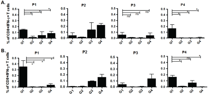
| Figure 6: Intra-cellular Cytokine Staining for CD8+/IFN-γ+ T cells after DNA-DNA immunization. 107 splenocytes were plated in 6 well plates and stimulated with relevant peptides (P1-P4). 3 days later, cell cultures were supplemented with rh.IL-2 at 100U/ml final concentration and incubated for further 3 days. Cells were harvested and restimulated in vitro again by relevant peptides, irrelevant control and PMA/Ion along with Golgiplug while incubated at 37°C and 5% CO2 incubator overnight. Cells were further stained for CD3/CD8/IFN-γ and data was acquired by BD FACScalibur on 100,000 events and analyzed by FlowJo software. A. 3 weeks after challenge. B. 7 weeks after challenge. Each column represents mean+SD of CD8+/IFN-γ+ T cells in each group. Significant differences are presented by stars after analysis by Mann-Whitney assay (0.05>p>0.01). ns: non-significant difference. |