|
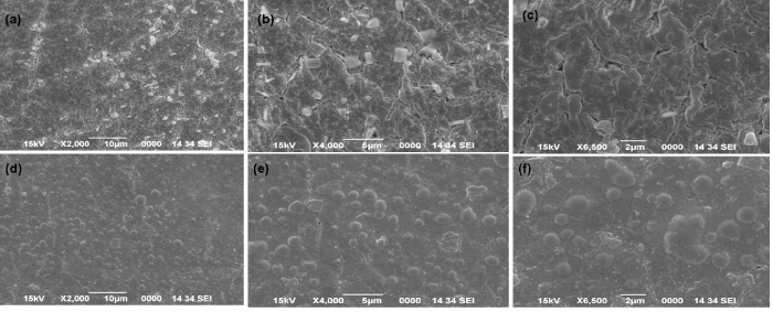
| Figure 6: Scanning electron micrographs obtained for the blank PAni-GA-sodium alginate and PAni-GA-sodium alginate-D-aao beads. (a), (b),(c) are the SEM images for the blank beads at different magnifications while (d), (e), (f) are the SEM images of the D-aao immobilized beads at different magnifications. |