|
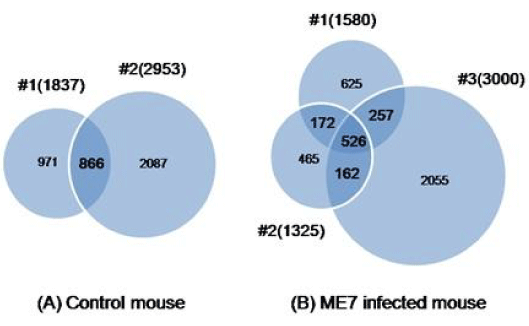
| Figure 3: Venn diagram showing the number of unique and shared proteins from each individual mouse. Venn diagram of the proteins identified by one-dimensional gel electrophoresis-liquid chromatography-tandem mass spectrometry (1D-Gel-LC-MS/MS) in ME7 scrapie-infected and control mice. (A) Two control mice (#1, #2). (B) Three ME7 scrapie-infected mice (#1, #2, #3). |