Taishi Okada, Hiroyuki Futani, Ryo Kanto, Shunsuke Kumanishi, Yoshitane Tsukamoto and Shinichi Yoshiya
Chondromyxoid fibroma is an extremely rare, benign cartilaginous tumor, which might be misdiagnosed as hondrosarcoma. Recent studies reported that PET/CT could distinguish benign cartilaginous tumors from chondrosarcomas with maximum Standardized Uptake Value (SUVmax) of more than 2.0. In the literature, 4 cases of chondromyxoid fibroma have been reported on PET/CT with high accumulation of 18FFDG. However, no paper has explained the reason for this high accumulation. In this paper, we present a case of femoral chondromyxoid fibroma and discuss the rational reason for high accumulation of 18F-FDG by PET/CT in accordance with histology. Here, a 20-year-old female presented with a lesion located in the medial aspect of the left distal femur. Radiography revealed an eccentric radiolucency in the metaphysis of the left distal femur. CT images clearly demonstrated a cortical destruction of the posterior wall. PET/CT images clearly demonstrated an abnormal 18F-FDG uptake of the distal aspect of the left femur with SUVmax value of 6.6, indicating a chondrosarcoma. In the present case, histology showed a number of multinucleated giant cells at the periphery of the lobules in the tumor, which can explain the high accumulation.
PDFShare this article
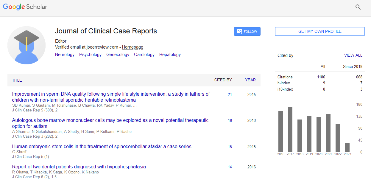
Journal of Clinical Case Reports received 1295 citations as per Google Scholar report