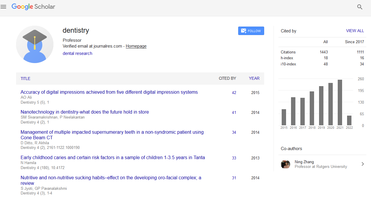
Citations : 1817
Dentistry received 1817 citations as per Google Scholar report
Indexed In
- Genamics JournalSeek
- JournalTOCs
- CiteFactor
- Ulrich's Periodicals Directory
- RefSeek
- Hamdard University
- EBSCO A-Z
- Directory of Abstract Indexing for Journals
- OCLC- WorldCat
- Publons
- Geneva Foundation for Medical Education and Research
- Euro Pub
- Google Scholar
Useful Links
Share This Page
Journal Flyer

Open Access Journals
- Agri and Aquaculture
- Biochemistry
- Bioinformatics & Systems Biology
- Business & Management
- Chemistry
- Clinical Sciences
- Engineering
- Food & Nutrition
- General Science
- Genetics & Molecular Biology
- Immunology & Microbiology
- Medical Sciences
- Neuroscience & Psychology
- Nursing & Health Care
- Pharmaceutical Sciences
Abstract
Relationship between Clinical and Histopathologic Findings of 40 Periapical Lesions
Francisco Javier Jimenez Enriquez, Jorge Paredes Vieyra and Fabian Paredes Ocampo
Purpose: To relate the clinical and histopathological findings of periapical inflammatory lesions treated by endodontic surgery with the results of histopathological investigation of the same lesions.
Materials and methods: Forty biopsies obtained during periapical surgery were histologically analyzed following curettage of the tissue, establishing the diagnosis as either periapical granuloma, radicular cyst, or abscess. The radiographic size of the lesion (area in cm2) before surgery and after 2 years of follow-up was measured. The evolution at 48 months after surgery was evaluated according to the criteria of von Arx and Kurt. A statistical study was made, the inter-variable relationships were studied using analysis of variance with subsequent Tukey testing and calculation of Pearson?s correlation coefficient. The hypothesis tests were conducted at the 0.05 level of significance.
Results: Results indicated that 26 (65%) were women and 14 (35%) men at a mean age of 43.54 years (range,18 to 69 years) with 40 biopsy samples. 65.5% of lesions were granulomas, 20% cysts, and 17.5% abscess. The results showed that tooth lower second molar had a high percentage of periapical lesion corresponding to periapical granuloma associated with overfilled canals.
Conclusions: The outcomes of the present study show a high number of periapical granulomas among periapical cysts and confirms that periapical granulomas are the most common periapical lesions of endodontic origin associated with persistent apical periodontitis.


