Sihem Darouich, Nadia Boujelb�?¨ne, Souhir Bouzguenda, Dhikra Kacem, Radhia Ben Ghorbel, Karima Mrad and Aida Masmoudi
University of Tunis El Manar, Tunisia
Posters & Accepted Abstracts: J Clin Case Rep
Hypophosphatasia is a rare skeletal dysplasia, related to inactivating mutations in the TNSALP gene encoding the tissue-nonspecific alkaline phosphatase. This enzyme deficiency mainly results in disorders of bone and teeth mineralization. The phenotypic spectrum ranges from perinatal lethal to mild form diagnosed in adulthood. We describe the lethal form in three affected fetuses, one female and two males. Antenatal ultrasound detected micromelia and generalized demineralization. Termination of pregnancy was performed at 18, 22 and 25 weeks of amenorrhea. Skeletal radiographs showed irregular ossification in all cases, that was very poor with only few mineralized bones in two cases, and associated with bowing of the long bones and large, irregular metaphyses in one case. Macroscopic examination showed micromelia, bowing of the limbs and a characteristic craniofacial dysmorphism including relative macrocrania, hypertelorism, small nose with anteverted nostrils, long philtrum, thin lips, miroretrognathia and low-set ears. Bilateral tibial or tibio-peroneal spurs were observed in two cases. The histology disclosed the presence of columns of hypertrophic chondrocytes invading the diaphysis. In conclusion, perinatal hypophosphatasia is characterized by severe micromelia and major deficit or complete absence of the bone mineralization. It is often confused with osteogenesis imperfecta on prenatal ultrasound. However, the 3D ultrasound can detect the characteristic bone spurs in the early second trimester and strongly orient the diagnosis towards lethal hypophosphatasia. The diagnosis is confirmed by detailed fetopathological examination including skeletal radiography, macroscopic examination and histological study of longs bones. Genetic counseling can be offered with genetic testing to parents.
Sihem Darouich, MD, is a Fetopathologist at the University Hospital Habib Bougatfa of Bizerte and Professor at the Department of Histology and Embryology, Faculty of Medicine of Tunis, Tunisia. Over the last several years, she has focused on fetal genetic syndromes, especially syndromic multicystic renal dysplasia and skeletal dysplasias. She is a member of the French Society of Fetopathology (SoFFoet) and of an editorial board of a reputed international journal.
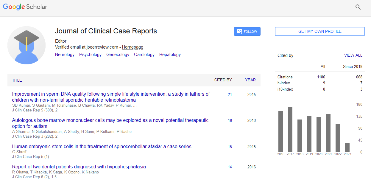
Journal of Clinical Case Reports received 1295 citations as per Google Scholar report

 Spanish
  Chinese
  Russian
  German
  French
  Japanese
  Portuguese
  Hindi
Hindi 